
Cardiac magnetic resonance (CMR) is a non-invasive imaging technique that provides detailed images of your heart’s structure and function. It is a powerful diagnostic tool used by healthcare professionals to identify and monitor various cardiovascular conditions. CMR uses strong magnetic fields and radio waves to capture high-resolution images of the heart, allowing for a thorough assessment of its anatomy, blood flow, and overall health. This advanced imaging technology plays a crucial role in the early detection and management of heart diseases, enabling healthcare providers to make more informed treatment decisions.
What is Cardiac Magnetic Resonance (CMR)?
Cardiac magnetic resonance (CMR) is a non-invasive medical imaging technique that uses strong magnetic fields and radio waves to create detailed, three-dimensional images of the heart. Unlike traditional X-ray or CT scans, CMR does not use ionizing radiation, making it a safer option for patients. The technology works by aligning the protons in the body’s hydrogen atoms with a strong magnetic field. Radio waves are then applied, causing the protons to absorb and release energy, which is detected by the scanner and translated into high-resolution images of the heart’s structure and function.
The principles of CMR imaging involve the use of a powerful magnetic field that interacts with the hydrogen protons in the body. When the patient is placed inside the CMR scanner, the magnetic field aligns the protons, and then radio waves are applied. As the protons absorb and release energy, the scanner detects these changes and transforms them into detailed, three-dimensional images of the heart. This non-invasive process allows healthcare providers to gain a comprehensive understanding of how the heart is functioning, enabling them to make more informed diagnostic and treatment decisions.
| Key Features of Cardiac Magnetic Resonance (CMR) | Explanation |
|---|---|
| Non-Invasive Imaging | CMR does not require any surgical or invasive procedures, making it a safe and comfortable option for patients. |
| Detailed Anatomical Information | CMR provides high-resolution, three-dimensional images of the heart’s structure, including the chambers, valves, and blood vessels. |
| Functional Assessment | CMR can evaluate the heart’s pumping ability, blood flow, and other important functional parameters. |
| No Ionizing Radiation | Unlike X-rays and CT scans, CMR does not use ionizing radiation, reducing the risk of potential side effects. |
The Role of CMR in Diagnosing Heart Diseases
Cardiac magnetic resonance (CMR) is a versatile diagnostic tool that plays a crucial role in the detection and management of various heart conditions. By providing detailed, non-invasive images of the heart, CMR allows healthcare providers to accurately diagnose and monitor a wide range of cardiovascular issues, including:
- Coronary artery disease – CMR can identify blocked or narrowed arteries, assess the extent of damage, and evaluate the heart’s function.
- Heart valve problems – CMR can accurately measure valve function, detect structural abnormalities, and guide treatment decisions.
- Cardiomyopathy – CMR is highly effective in diagnosing and characterizing different types of cardiomyopathy, such as hypertrophic, dilated, and restrictive forms.
- Congenital heart defects – CMR provides detailed images of complex congenital heart structures, helping physicians plan appropriate interventions.
- Myocarditis and pericarditis – CMR can detect inflammation in the heart muscle and surrounding tissues, enabling early diagnosis and treatment.
- Aortic disease – CMR can accurately assess the size, shape, and function of the aorta, identifying dissections, aneurysms, and other aortic abnormalities.
By leveraging the advanced imaging capabilities of CMR, healthcare providers can better understand the underlying causes of heart problems, monitor disease progression, and make more informed treatment decisions. The role of CMR in diagnosing heart diseases continues to grow, as this non-invasive technology becomes an increasingly valuable tool in the field of cardiology.
Advantages of CMR Over Traditional Imaging
Cardiac magnetic resonance (CMR) offers several distinct advantages over traditional imaging techniques like X-ray, CT scans, and echocardiography, making it a superior choice for diagnosing and managing cardiovascular conditions. Unlike X-ray and CT scans, CMR does not use ionizing radiation, making it a safer option, especially for patients who require frequent follow-up imaging. Additionally, CMR provides detailed, high-resolution images of the heart’s structure and function, allowing healthcare providers to make more informed treatment decisions.
One of the key advantages of CMR is its ability to visualize the heart’s soft tissues in great detail. This allows clinicians to identify and monitor a wider range of cardiovascular issues, including myocardial infarction, cardiomyopathy, and valvular heart disease, that may not be as easily detected by other imaging techniques. Furthermore, CMR can assess blood flow and cardiac function with exceptional accuracy, providing vital information for the management of heart conditions.
Compared to echocardiography, CMR offers superior image quality and the ability to visualize the entire heart, including hard-to-reach areas. This comprehensive assessment can lead to more accurate diagnoses and the identification of subtle changes in cardiac structure and function that may be missed by other imaging modalities. Additionally, CMR is not limited by factors such as body habitus or poor acoustic windows, making it a valuable tool for evaluating patients who may not be suitable candidates for echocardiography.
| Imaging Technique | Advantages of CMR |
|---|---|
| X-ray |
|
| CT Scan |
|
| Echocardiography |
|
In summary, the advantages of cardiac magnetic resonance (CMR) over traditional imaging techniques make it a valuable diagnostic tool in the field of cardiology. By providing detailed, non-invasive images of the heart and its functions, CMR enables healthcare providers to make more accurate diagnoses and develop more effective treatment plans for their patients.
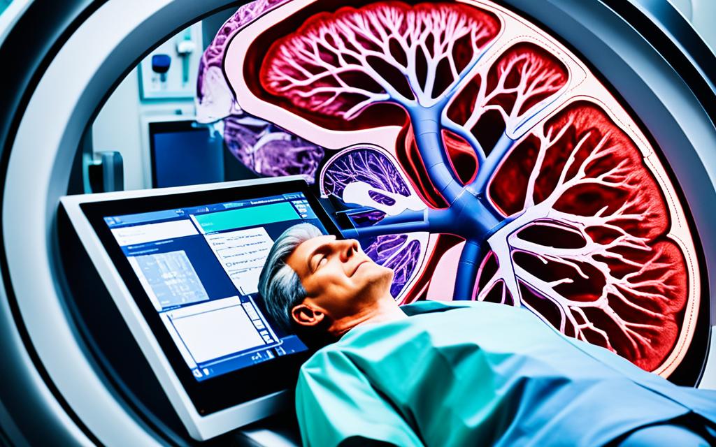
Cardiac Magnetic Resonance: What to Expect
If your healthcare provider has recommended a cardiac magnetic resonance (CMR) scan, it’s important to understand what to expect during the procedure. Here’s a step-by-step overview of the CMR scan process:
Preparation: Before your CMR scan, you may be asked to fast for a few hours, avoid caffeine, and wear comfortable clothing without metal fasteners or zippers. This helps ensure the best possible images and minimizes any potential interference during the scan.
The Scanning Process: When you arrive for your CMR scan, you’ll be asked to lie down on a movable table that slides into the scanner, which is a large, cylindrical machine. The technician will position you and may ask you to hold your breath briefly at various points during the scan to obtain clear images. The CMR scan procedure is generally painless, but you may experience a slight feeling of confinement or claustrophobia while inside the scanner.
| Step | Description |
|---|---|
| Preparation | Fasting, avoiding caffeine, and wearing comfortable clothing without metal |
| Positioning | Lying down on a movable table that slides into the CMR scanner |
| Imaging | Holding your breath briefly at various points to obtain clear images |
The CMR scan typically takes 30 to 60 minutes to complete, depending on the specific tests your healthcare provider has ordered. Throughout the process, the technician will be monitoring you and providing instructions as needed.
After your CMR scan, you can typically resume your normal activities. Your healthcare provider will review the images and provide you with the results, which can help them make a more informed diagnosis and develop an appropriate treatment plan for your condition.
Conclusion
As the field of cardiology continues to evolve, the role of cardiac magnetic resonance (CMR) imaging in the detection, monitoring, and management of cardiovascular conditions is poised to grow even more prominent. The latest advancements in CMR technology have expanded its capabilities, allowing healthcare providers to gain unprecedented insights into the structure and function of the heart.
The future of CMR in cardiology is bright, with researchers and clinicians exploring new applications to improve heart disease diagnosis and treatment. From the early identification of subtle cardiac abnormalities to the precise tracking of disease progression, CMR is revolutionizing the way healthcare professionals approach cardiovascular care.
By providing non-invasive, detailed images of the heart, CMR is helping clinicians make more informed decisions, leading to better patient outcomes. As this cutting-edge technology continues to advance, you can expect to see CMR play an increasingly vital role in the comprehensive management of a wide range of heart conditions, from congenital defects to ischemic heart disease.