
Computer tomography (CT), also known as computerized axial tomography (CAT) scans, are a type of medical imaging technology that creates detailed, cross-sectional images of the body. These scans use X-rays and computer processing to produce high-quality images that can help healthcare providers diagnose and monitor a wide range of medical conditions. In this comprehensive guide, you’ll learn about the technology behind CT scans, their various uses in healthcare, what to expect during the procedure, and the potential risks and benefits of this important diagnostic tool.
What is Computer Tomography (CT)?
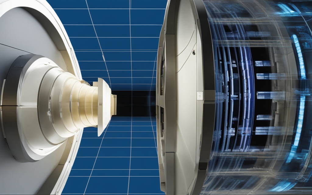
Computer tomography (CT), also known as computerized axial tomography (CAT) scans, is a medical imaging technique that uses X-rays and computer processing to create detailed, three-dimensional images of the inside of the body. During a CT scan, you lie on a table that slides into a large, donut-shaped machine. The machine rotates around your body, taking multiple X-ray images from different angles. A computer then assembles these images into cross-sectional views, or “slices,” of the area being scanned. These detailed images can reveal information about the size, shape, and position of organs, tissues, and other structures within the body.
The CT scan process allows healthcare providers to closely examine the internal structures of the body, enabling more accurate diagnoses and better-informed treatment decisions. By understanding how computer tomography works, you can be better prepared for your upcoming scan and work closely with your medical team to address any concerns or questions you may have.
| Key Features of CT Scans | Benefits of CT Scans |
|---|---|
| Uses X-rays and computer processing to create 3D images | Provide highly detailed images of organs, tissues, and structures |
| Rotates around the body, taking multiple X-ray images | Help diagnose a wide range of medical conditions |
| Assembles the images into cross-sectional “slices” | Guide treatment decisions and monitor the effectiveness of therapies |
By understanding the fundamentals of computer tomography and how CT scans work, you can be better prepared for your upcoming medical imaging procedure and have a more informed discussion with your healthcare provider.
How CT Scans Work
The process of creating a CT scan image begins with an X-ray tube located inside the CT scanner. The X-ray tube rotates around your body, taking multiple images from different angles. As the X-rays pass through your body, they are absorbed and scattered in different ways depending on the density of the tissues they encounter. The CT scanner’s detectors measure the X-ray attenuations, and a computer uses this information to reconstruct a detailed, three-dimensional image of the scanned area. The resulting images are displayed on a monitor, allowing healthcare providers to closely examine the internal structures of the body.
The key steps in the CT scan process are:
- The X-ray tube inside the CT scanner rotates around the patient’s body, taking multiple X-ray images from different angles.
- As the X-rays pass through the body, they are absorbed and scattered in different ways, depending on the density of the tissues.
- The CT scanner’s detectors measure the X-ray attenuations, or the changes in the X-ray beam as it passes through the body.
- A computer uses the attenuation data to reconstruct a detailed, three-dimensional image of the scanned area.
- The resulting images are displayed on a monitor, allowing healthcare providers to closely examine the internal structures of the body.
The CT scan technology used in modern CT scanners is highly advanced, allowing for faster scan times, lower radiation doses, and higher-quality images than ever before. By understanding how CT scans work, you can better appreciate the role this important imaging technique plays in modern healthcare.
Uses of CT Scans in Medicine
CT scans have become an invaluable tool in the healthcare industry, allowing medical professionals to diagnose and monitor a wide range of medical conditions. From identifying cancerous tumors to detecting cardiovascular diseases, these advanced imaging techniques are playing a crucial role in modern medicine.
One of the primary uses of CT scans is in the diagnosis and staging of cancer. These scans can detect the presence of tumors and provide detailed information about their size, shape, and location, which is essential for developing an effective treatment plan. Additionally, CT scans can be used to monitor the effectiveness of cancer treatments, helping healthcare providers track the progress of the disease and make informed decisions about the next steps in a patient’s care.
Another important application of CT scans is in the realm of cardiovascular disease. These scans can reveal blockages in the arteries, providing valuable insights that can help healthcare providers diagnose and treat heart conditions. By identifying areas of concern, such as narrowed or blocked blood vessels, CT scans can guide the development of targeted treatment strategies, including surgical interventions or medication management.
In cases of trauma, CT scans play a crucial role in quickly identifying injuries to the head, chest, and abdomen. These scans can detect internal bleeding, organ damage, and other life-threatening conditions, allowing healthcare providers to initiate timely and appropriate treatment, ultimately improving patient outcomes.
CT scans are also valuable in the diagnosis and monitoring of infections, particularly in the lungs, sinuses, and other parts of the body. By providing detailed images of the affected areas, these scans can help healthcare providers identify the source of an infection and develop an effective treatment plan.
Finally, CT scans are widely used to assess skeletal issues, including fractures, degenerative changes, and other problems affecting the bones, joints, and spine. The detailed images generated by these scans can help healthcare providers accurately diagnose and monitor these conditions, guiding treatment and rehabilitation efforts.
Overall, the uses of CT scans in medicine are vast and ever-expanding, with these advanced imaging techniques playing a crucial role in the diagnosis, management, and monitoring of a wide range of medical conditions. As technology continues to evolve, the potential of CT scans to transform healthcare and improve patient outcomes is likely to grow even further.
Preparing for a CT Scan
Before your CT scan, you may need to take specific steps to prepare. It’s important to follow all instructions provided by your healthcare team to ensure the scan is as accurate and effective as possible.
One common preparatory step is fasting. You may be asked to avoid eating or drinking for several hours before the scan, especially if the scan will include the abdomen or pelvis. This helps ensure optimal image quality.
You may also need to drink a contrast medium, such as a barium-based solution, to help highlight certain areas of your body during the CT scan. In some cases, you may receive an intravenous contrast dye injection before the scan to help certain structures show up more clearly.
Additionally, your healthcare provider may give you specific instructions about taking or avoiding certain medications before the CT scan procedure. It’s important to follow these guidelines carefully to avoid any potential interactions or complications.
| Preparation Step | Purpose |
|---|---|
| Fasting | Ensure optimal image quality, especially for scans of the abdomen or pelvis |
| Drinking contrast medium | Highlight specific areas of the body for improved visualization during the CT scan |
| Intravenous contrast dye | Help certain structures show up more clearly in the CT scan images |
| Medication instructions | Avoid potential interactions or complications during the CT scan procedure |
By following the instructions provided by your healthcare team, you can help ensure that your CT scan is as accurate and informative as possible, ultimately supporting your overall health and wellbeing.
Computer Tomography
Computer tomography (CT), also known as computerized axial tomography (CAT), is a medical imaging technique that uses X-rays and computer processing to create detailed, three-dimensional images of the inside of the body. During a CT scan, the patient lies on a table that slides into a large, donut-shaped machine. The machine rotates around the patient’s body, taking multiple X-ray images from different angles. A computer then assembles these images into cross-sectional views, or “slices,” of the scanned area. These detailed images can reveal information about the size, shape, and position of organs, tissues, and other structures within the body, helping healthcare providers diagnose and monitor a wide range of medical conditions.
The CT scan process involves the use of advanced CT scan technology, which utilizes X-rays and computer processing to generate highly accurate and informative images of the body’s internal structures. These images can provide valuable insights into a patient’s health, assisting healthcare professionals in the diagnosis and treatment of various medical conditions, from cancer and cardiovascular disease to skeletal issues and infections.
By understanding the capabilities of computer tomography, you can work closely with your healthcare team to determine if a CT scan is the appropriate diagnostic tool for your specific medical needs. With its ability to capture detailed, three-dimensional images of the body, CT scans continue to play a vital role in modern healthcare, helping to improve patient outcomes and enhance the overall quality of medical care.
Risks and Benefits of CT Scans
Like any medical procedure, CT scans come with both risks and benefits that patients should be aware of. Understanding these factors can help you make an informed decision about whether a CT scan is the right choice for your specific healthcare needs.
On the risks side, the primary concern with CT scans is the exposure to a small amount of ionizing radiation. This radiation can potentially increase the risk of cancer over time, especially with repeated scans. Additionally, some patients may experience an allergic reaction to the contrast dye used in certain CT scans.
However, the benefits of CT scans often outweigh these risks. These advanced imaging techniques provide healthcare providers with highly detailed, three-dimensional images that can improve diagnosis and lead to faster, more effective treatment. In some cases, CT scans can even reduce the need for more invasive procedures, such as surgery.
Ultimately, it’s important to discuss the potential risks and benefits of CT scans with your healthcare provider. They can help you determine if a CT scan is the best course of action for your specific medical needs and ensure the safety of the procedure.
What to Expect During and After a CT Scan
During a CT scan, you can expect to lie on a table that slides into the CT scanner. You’ll need to remain still and hold your breath briefly as the scanner rotates around you. Depending on the type of scan, you may also receive an injection or need to ingest a contrast dye to enhance the images.
After the CT scan procedure, there is typically no significant recovery time, as it is a non-invasive test. However, you may experience minor side effects from the contrast dye, such as nausea or a warm sensation. It’s important to follow any post-scan instructions provided by your healthcare team and to ask questions if you have any concerns or experience any unusual symptoms following the procedure.
Once the CT scan is complete, your healthcare provider will review the detailed images and discuss the findings with you, as well as the next steps in your care. This open communication is essential for ensuring you understand the results and feel informed about your treatment plan.
| What to Expect During a CT Scan | What to Expect After a CT Scan |
|---|---|
|
|
By understanding what to expect during and after a CT scan, you can feel more prepared and confident throughout the process. Remember to follow any instructions provided by your healthcare team and don’t hesitate to ask questions if you have any concerns.
Conclusion
In conclusion, computer tomography (CT) scans are a valuable diagnostic tool that provide healthcare providers with detailed, three-dimensional images of the inside of the body. These scans can help identify a wide range of medical conditions, from cancer and heart disease to infections and skeletal issues. While there are some potential risks associated with CT scans, such as radiation exposure, the benefits often outweigh the risks, especially for time-sensitive or complex medical concerns.
By understanding the CT scan process, what to expect before, during, and after the procedure, and the potential risks and benefits, you can work closely with your healthcare team to determine if a CT scan is the right choice for your medical needs. This comprehensive guide has provided you with the key takeaways about computer tomography to help you make an informed decision about your healthcare.
Remember, your health and well-being are the top priorities, and your healthcare team is there to guide you through the process and ensure you receive the best possible care. If you have any further questions or concerns, don’t hesitate to discuss them with your doctor or healthcare provider.